

3D Dental Imaging Technology in Edinburg, TX
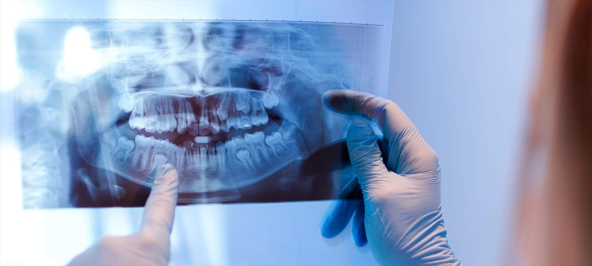
i-CAT FLX Imaging System
The i-CAT FLX is a Cone Beam Volumetric Tomography and Panoramic Imaging System used for dental head and neck applications. It consists of a scanner, scanner controller, touch screen, and keyboard which is suitable for an in-office environment.
The scanner is an open design that allows patients to sit upright during a procedure. An electric-powered seat is built into the scanner for proper patient positioning.
Cone Beam Volumetric Tomography is a medical imaging technique that uses X-rays to obtain cross-sectional images of the head or neck. The quality of the images depends on the level and amount of X-ray energy delivered to the tissue.
Imaging displays both high-density tissue, such as bone, and soft tissue. When interpreted by a trained physician, these images provide useful diagnostic information.
The scanner captures data for 3D skull reconstruction for the following procedures:
- Implants
- TM Joints
- Reconstructed Panoramic
- Reconstructed Cephalometric
- Airway / Sinus, etc.
- Nerve Canal
- PAN – Optional Conventional Digital Panoramic Feature
We’re Here To Help!
Call us with any questions or to schedule an appointment.